Associate Professor of Hematology, Consultant of Medical Laboratory Sciences, Port Sudan Ahlia University, Faculty of Medical Laboratory Sciences, Port Sudan, Sudan.
Bashir Abdrhman Bashir
Tel: 00249912358772, Fax: 00249 3118 26537;
Email: bashirbashir17@hotmail.com
Received : Aug 04, 2025 Accepted : Sep 07, 2025 Published : Sep 14, 2025 Archived : www.meddiscoveries.org
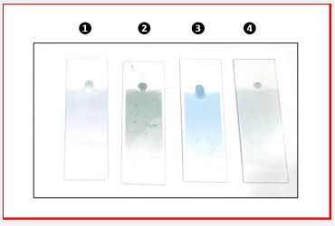
This image presents four supravital-stained blood films for the assessment of reticulocytes utilizing fresh methylene blue.The slides exhibit distinct backdrop colors, each signifying a specific diagnostic or technical implication:
Slide 1 (Pale Blue): The staining issue, perhaps attributable to inadequate dye concentration or fast drying, resulting in suboptimal reticulocyte visibility (Reticulocyte count 4.3%, Hb A).
Slide 2 (Green Hue): This coloring may result from an alkaline pH change, potentially due to elevated amounts of fetal Hemoglobin (HbF), as shown in β-thalassemia, hereditary persistence of HbF, or during neonatal erythropoiesis. It may also indicate inadequate buffering or oxidized stain (Reticulocyte count 19.1%, Hb E & B-thal).
Slide 3 (Bright Blue): Illustrates optimal staining conditions characterized by ideal pH and stain activity, resulting in distinctly recognizable reticulocytes with clearly defined granules (Reticulocyte count: 3.7%, Hb A).
Slide 4 (Clear/Central Drop): Signifies inadequate mixing or drying artifact; reticulocyte distribution may be irregular, undermining accuracy (Reticulocyte count 10.2%, Hb AS).
Reticulocyte labeling, a powerful tool in our quest to understand the biology of erythropoiesis, enables the direct observation of immature erythrocytes by the precipitation of leftover RNA using supravital dyes. In addition to reticulocyte counting, the background color serves as a prompt indicator of staining quality and possible hematological disorders, offering a constant intellectual challenge.
The greenish hue, often dismissed as a technical defect, demands careful consideration. It necessitates an examination of biological factors like increased HbF, which modifies intracellular buffering and may react differently with the stain, particularly in neonates or thalassemic individuals. In certain situations, the alkaline intracellular milieu alters the stain’s optical characteristics, resulting in a distinctive greenish tint that can significantly impact our interpretation of results.
Neglecting to acknowledge the subtleties of background staining may lead to Inaccurate reticulocyte counts, Misinterpretation of hematopoietic function, and Oversight of illnesses such as β-thalassemia major, HbF abnormalities, or pH-sensitive enzymatic anomalies. Consequently, background examination ought to be integrated as a standard component of morphological quality control, especially in contexts assessing juvenile anemia, hemoglobinopathies, and bone marrow response.
This image emphasizes the essential yet frequently disregarded diagnostic significance of background coloration in reticulocyte labeling. A green stain background may not solely signify a technical fault but could also suggest biological changes, such as elevated HbF levels or alkaline pH conditions. The integration of morphological observation with clinical context improves diagnosis accuracy in hematology.